Introduction
Background
Anatomical study
History
Dynamic tip
Bridge support
Photo planning
Operation
Results
Publications
|
Anatomical Study
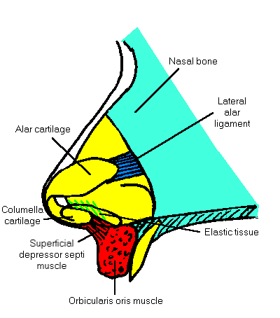
Paul O'Keeffe investigated the anatomy of the columella base
and membranous septum during 1989. The study was carried out at
the Sydney Morgue in Glebe.
| Young cadavers were found to have on each
side a relatively large pyramidal-shaped
muscle arising from a fibrous layer on the
anterior surface of the orbicularis oris muscle
and inserting into the medial surface of the ipsilateral
footplate of the columella cartilage. This
muscle may be called the superficial depressor septi
muscle. The muscle appeared to be smaller in
older cadavers who had a short distance between the foot
plates and the orbicularis oris muscle. The muscle would appear
to be the functional tether between the columella and the
lip. |

 |
| The membranous septum was resected and
examined histologically. It was stained for elastin revealing a
rich elastic fibre content. The elastic fibres
are arranged primarily in one direction and their orientation
would protrude the columella and nose tip after retrusion caused
by contraction of the superficial depressor septi muscle. The
elasticised mucous membrane covers the caudal margin of the
septal cartilage. |

Elastic fibres are stained
black
|

Back to Top
|


